Teleradiology has been used for years in both human and veterinary medicine. When we think of “teleradiology,” we usually consider evaluation of electronically stored and transmitted radiographs, CT or MRI by a boarded radiologist, who can be anywhere in the world that has internet access. The advantages are numerous and include rapid access to boarded specialists that otherwise would not be possible. Telemedicine, on the other hand, is relatively new, but is rapidly expanding and offers similar access to other specialists. When we think of telemedicine, there are certain specialties that are more amenable to “distant” medical practice than others. Medical specialties that require hands-on type work, such as surgery, make it difficult (but not impossible with robotics). However, telemedicine, teledermatology, teleophthalmology, etc. are all possible and we are starting to see the beginnings of these other specialties now.
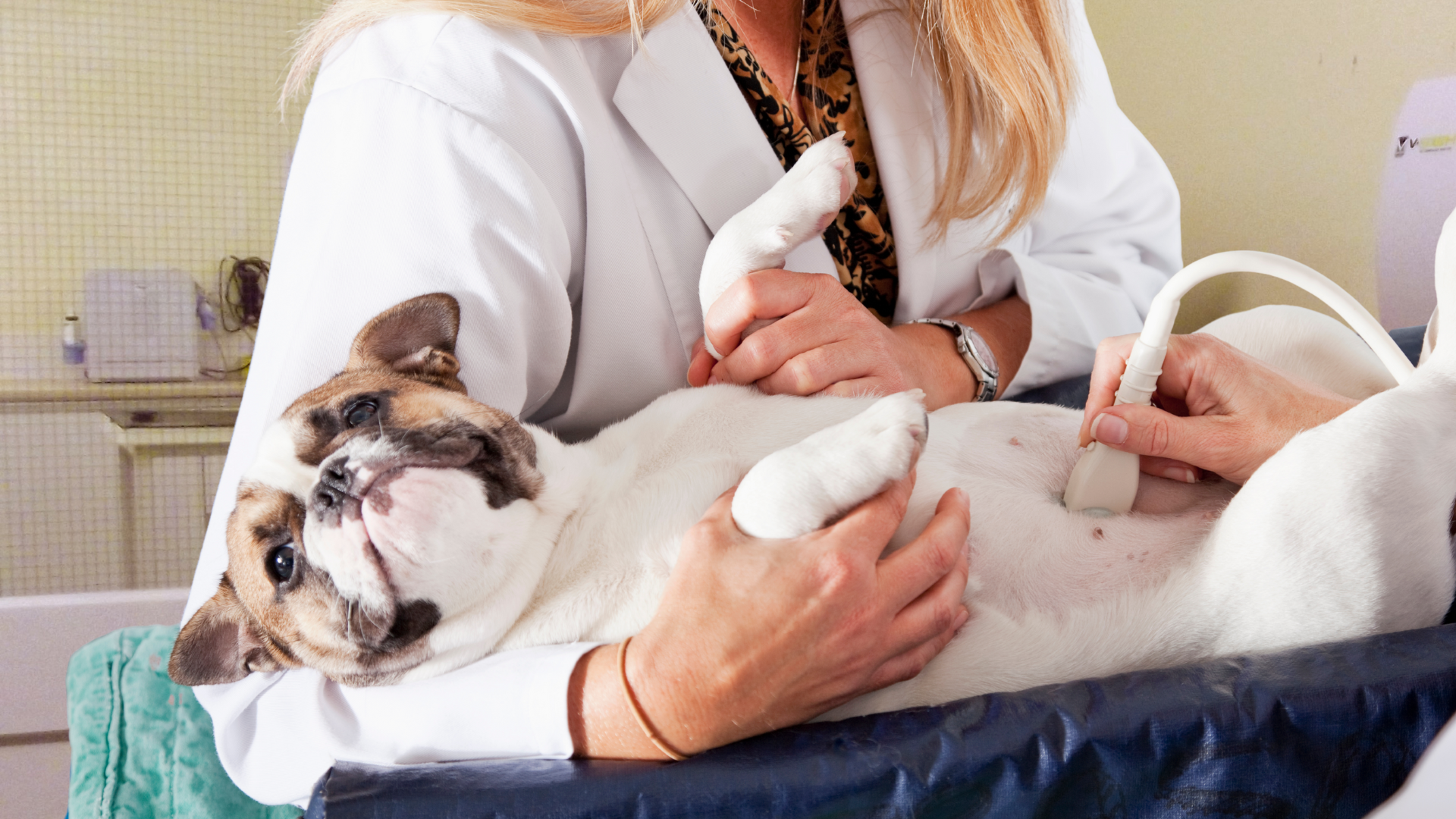
Teleultrasound has long been the odd-ball when it comes to remote imaging and interpretation, because it is also highly dependent on the hands-on, “probe-in-hand,” physical ability to scan and find organs and obtain quality images of what you see. Because of these limitations, there are certain radiologists that will NOT read anyone else’s ultrasound exam. VitalRads policy is different, and if the following guidelines are followed, we have a lot of success reading remotely obtained ultrasound exams.
- The person obtaining the ultrasound images, must be well trained in VETERINARY ultrasound. Because of anatomic variances, human trained sonographer cannot jump right in and scan dogs and cats and will require additional training and practice in animals. Most veterinary sonographers get their training from attending didactic lectures on veterinary ultrasound, taught by radiologists, followed by months and years of practice. Interestingly, board certified veterinary internists commonly perform ultrasound exams in veterinary practices; however, they obtain their ultrasound training in the exact same way that a general veterinarian can learn ultrasound (i.e., they are not formally trained on ultrasound during their residencies). Regardless of the training, there is no substitute for hands-on scanning and practice. From VitalRads perspective, we can usually tell if the sonographer is competent or not by looking at the images that they submit.
- The typical abdominal ultrasound study submitted to us for evaluation consists of about 50-60 images, of which 90% are static images and a handful of short, 2-3 second video clips. Video clips should be obtained of any potential abnormalities/pathology seen during the exam. It is NOT necessary to obtain a video clip of a normal organ! It is also not necessary to obtain 30 images of the same normal organ.
- Keep in mind that as the radiologist, we will always be at a disadvantage because we do not have a probe in our hands and are relying on you to send us representative, labeled, correctly oriented, good quality images along with an appropriate history.
- A “private comment” from the sonographer about what their thoughts are when doing the scan and what they saw are very helpful in putting a “virtual” probe in our hands.