When it comes to providing accurate imaging reports, please keep the following “Top Ten List” in mind.
1. You cannot evaluate bowel wall thickness on plain films alone. Fluid within the bowel lumen silhouettes with the bowel wall (they are both “soft tissue equivalent” when it comes to radiology) causes the bowel wall to artifactually appear thickened. Ultrasound must be employed to confirm or deny suspect bowel wall thickening seen on radiographs.
2. “Whole body” films are NOT the way to go. Please do not fall into the “let’s just shoot the entire animal because you never know what you might see” attitude. This leads to non-diagnostic quality films. Please limit the field of view to the area of interest, based on the history, physical exam and orthopedic/neurologic exam findings. With the exception of some exotics, how many “whole body” films were shot at your veterinary school? None! There is a reason for this.
3. Lateral views that include BOTH right and left limbs on the same films will almost always result in non-diagnostic, poorly positioned films.
4. Positioning is critical especially for neurologic abnormalities.
5. Even slight obliquity on the lateral view will lead to underdiagnosis or misdiagnosis in the TL spine.
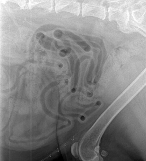
6. Cervical spines need to be “stretched,” straight, and the film needs to be centered over the spine.
7. Always center the film over the region of interest. For example, if the animal has cervical pain, center the films over the neck – not over the thorax, liver, or abdomen.
8. Always get the region of interest on the film! If the dog is vomiting, make sure and get the entire abdomen on the film. This sounds intuitive, but we often receive images that do not include the stomach or entire abdomen on patients with a history of vomiting.
9. Ultrasound is insensitive to judge the normal size of the liver or spleen. Radiographs are much more accurate.
10. CT is nearly always needed for abnormalities of the nasal, skull, or tympanic bulla. Even high quality and well positioned radiographs of these regions will underestimate pathology.
11. MRI is nearly always needed for suspect brain and spinal tumors. Even contrast CT is nowhere near as accurate as MRI.
12. The radiologist does not have magic tools to evaluate your films. An accurate reading is directly dependent on the provided history and film quality. Garbage in – garbage out.